26 Lab 8: Horizontal Slices & Case Study Development
Learning Objectives
- Review structures visible in midsagittal section to find landmarks for two horizontal dissection slices
- Make two horizontal dissection slices of the sheep brain
- Locate and identify structures within those two slices
- Use knowledge of drugs covered in Chapter 7 to create a case study
Horizontal Slices
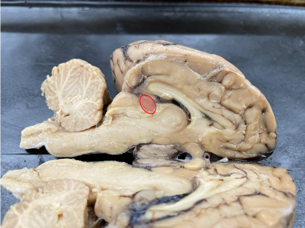
In this final dissection lab, you will be investigating the specimens in the horizontal or transverse plane. Familiarize yourself with identifying and/or locating these structures: 1) pineal body (See image below.) and 2) thalamus. You can use these structures to identify where you should position your scalpel to make each dissection slice. For the pineal body, review your work on the mid-sagittal slice. For the thalamus, you might review your work on coronal slices 3a and 3b.
Procedure:
- Place the hemisphere you selected for horizontal slices from your whole specimen in a dissection tray. Look at the midsagittal view, and locate the pineal body. Because it would be easier to cut the specimen while the mid-sagittal view is on the wax of the dissection tray, we suggest you use two pins to identify the anterior and posterior points of the brain you would need to cut to dissect in a straight line through the pineal body. In other words, decide on some markers on the lateral view that will allow you to cut through the pineal body, and place pins in those markers. Then, lay the specimen on its mid-sagittal side.
- Cut anterioposteriorly based on the markers you identified when looking at the pineal body.
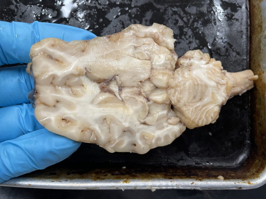
- After making your slice, you can view the structures on the inferior surface of that cut you made. This is horizontal slice 1 – the most superior dissection cut you will make during this lab.
REVIEW BOX
On the following image, and on your own slice, be sure you can identify these structures:
- Head of the caudate
- Lateral ventricle (Identifying the ventricle may be more easily accomplished with the use of a probe on your specimen than by looking at the image.)
- Cerebellum
- Corpus callosum
- A gyrus
- A sulcus
- Cortical gray matter
- White matter
- Septum pellucidum (While this structure is depicted in horizontal slice 1 of our resource books, it is not visible in the dissection slice depicted. Look to horizontal slice 2 image, instead.)
4. Next, considering what you know about the thalamus in relation to visible structures on the mid-sagittal slice or lateral view, select some markers that will allow you to cut through at about the middle of the thalamus, and use the pins as previously discussed (to help you envision a straight dissection line through the thalamus).
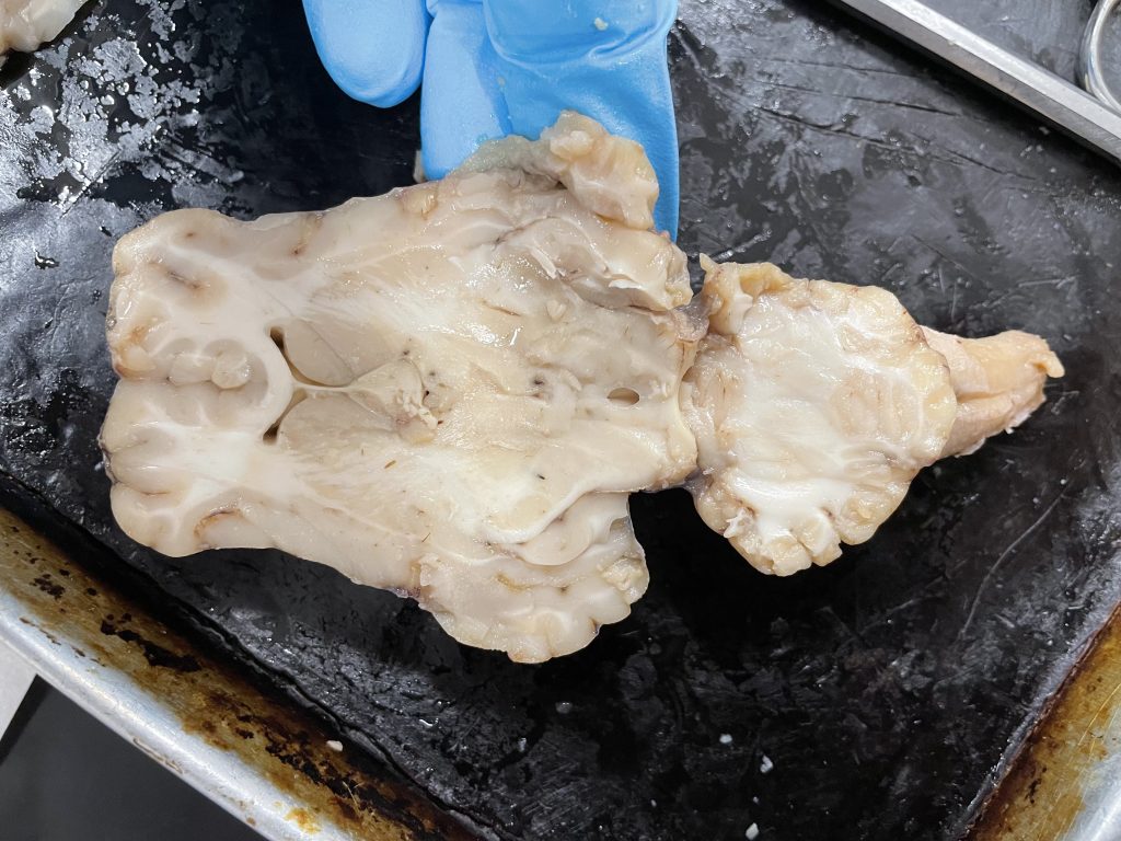
5. Next, cut anterioposteriorly along the imagined line connecting the markers you identified. The inferior surface is horizontal slice 2.
If you think it will help, view the video discussing these dissections. The video can be found in the Resources section of your lab Moodle site.
REVIEW BOX
On the following image, and on your own slice, be sure you can identify these structures:
- Lateral ventricle
- Cerebral aqueduct (in the image)
- Cerebellum
- Corpus callosum
- A gyrus
- A sulcus
- Cortical gray matter
- White matter
- Lateral geniculate nucleus (LGN)
- Medial geniculate nucleus (MGN)
Case Study Development
In groups of no more than 3, create one case study detailing a fictional character who is currently using the substance (one substance will be given to each table). In your case study, be sure to given a brief paragraph detailing the person’s demographics of relevance and the symptoms they are exhibiting. Then, create 2 or 3 questions (and provide answers) guiding someone who would read your case study to figure out which substance(s) are likely what your character is using. Then, develop 2-3 more sentences to help your reader distinguish between substance(s) if more than one substance could cause the symptoms or if there is any room for confusion. Then, in one sentence clarify which substance is the one being used. Finally, ask questions (and provide answers) to guide the reader through how that substance works in/on the human brain. (Consider what you learned in the chapter and in class. You don’t need to do outside research for this assignment.)
Your document should provide the names of all group members who helped develop the case study. Indicate whether you would be willing to allow your case study to be used in future labs or course materials and which license you would apply.
