19 Lab 1: Gross Anatomy
Erin Mazerolle and Sherry Neville-MacLean
Learning Objectives
- Understand and comply with lab safety rules
- Identify neuroanatomical directions and when they apply
- Identify neuroanatomical planes and when they apply
- Remove the dura mater
- Perform a midsagittal section
- Identify cortical brain lobes and other major structures visible from the surface and midsagittal plane
Lab Safety Rules
- Absolutely no eating or drinking in the lab. If you need to eat or drink during the 75 min lab period, you must leave the room to do so. There are absolutely no exceptions to this rule. Water bottles are not permitted in the lab.
- You must wear gloves and goggles while handling brain specimens.
- No sandals or other open footwear.
- Lab coats are recommended.
- Do not stick the dissection tools into the tray wax.
- You must clean up your materials after lab.
By participating in lab, you agree to comply with all the lab safety rules and any instructions of your instructor.
Materials
specimen
dissection tray
scissors
scalpel
forceps
probe
Removal of Dura Mater
This section adapted from Human Anatomy Laboratory Manual 2021 under a CC-BY license.
The sheep brain is enclosed in a tough outer covering called the dura mater. You can still see some structures on the brain before you remove the dura mater.

Take special note of the pituitary gland and the optic chiasma. These two structures will likely be pulled off when you remove the dura mater.

Remove the dura mater as instructed. (Your lab instructor or assistant will demonstrate at the beginning of the lab.)

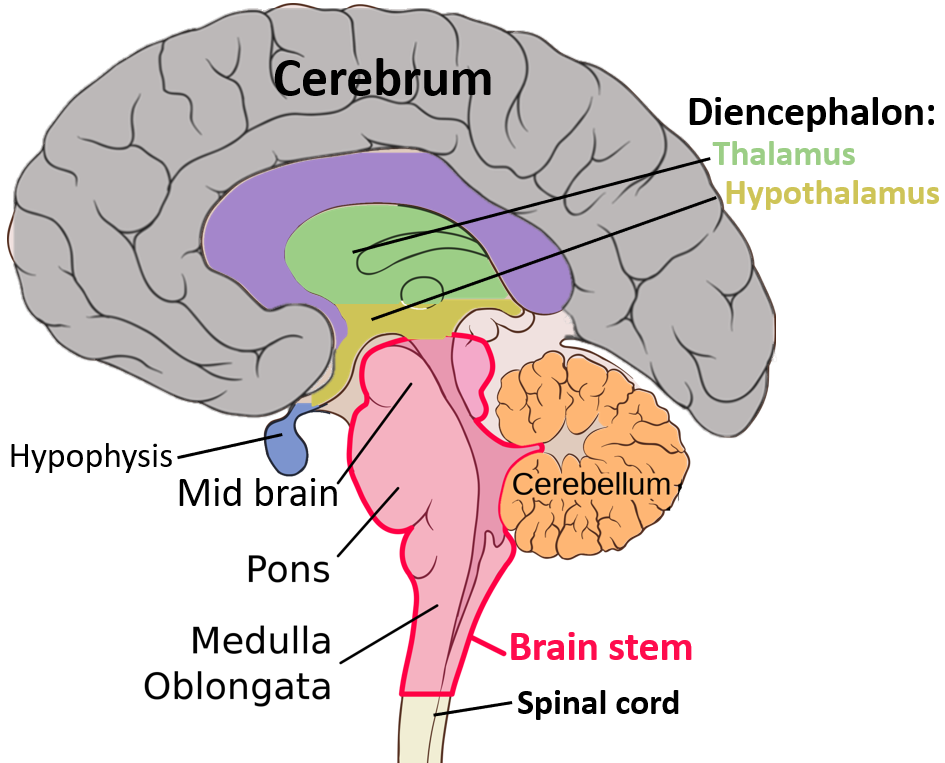
Neuroanatomical Directions of the Intact Brain + Cortical Brain Lobes, the Brainstem, and the Cerebellum
Hold and rotate your specimen. Look at the dorsal, ventral, and lateral views. Identify the right and left hemispheres, defined by the longitudinal fissure. Identify the cerebellum, separated from the cerebrum by the transverse fissure.
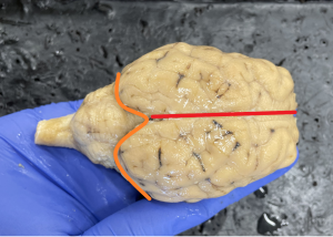
Identify the frontal, parietal, occipital, and temporal lobes. You are not expected to know the precise boundaries of most lobes of the sheep brain; however, you should be able to identify their general vicinity and location relative to each other. See a useful figure here.
Identify the brainstem.
Neuroanatomical Directions: Intact Brain
|
Term(s) |
Meaning |
|
Rostral/Anterior |
rostral – towards the nose anterior – towards the front |
|
Caudal/Posterior |
caudal – towards the tail posterior – towards the backside |
|
Dorsal/Superior |
dorsal – towards the back superior – above |
|
Ventral/Inferior |
ventral – towards the stomach/belly |
|
Lateral |
lateral – away from the midline; towards the extremities |
|
Medial |
medial – towards the midline |
Neuroanatomical Planes and Directions for Sections
Once we are “inside” the brain (i.e. sagittal or coronal slices or sections), we tend to use the terms superior and inferior to describe the position of structures rather than dorsal and ventral. Dorsal/ventral are used to describe the surfaces of the brain only.
Example:
|
Incorrect Terminology |
Correct Terminology |
|
The thalamus is dorsal to the hypothalamus. |
The thalamus is superior to the hypothalamus. |
Performing a Midsagittal Section
1. Place your specimen in a dissection tray with its dorsal surface up.
2. Be sure you have a pair of forceps and a scalpel ready at hand. A probe is also helpful to have nearby so that you can point to small structures when discussing them with your partner(s). Please use caution when handling the scalpels as they may be very sharp and do not get sterilized between uses.
3. When instructed, begin at the most anterior portion and place the scalpel blade along the medial longitudinal fissure. Draw the blade toward you in a single fluid motion. Do not plunge the blade into the specimen. If the blade becomes stuck (it hits a snag), do not re-cut the same area over, rather, spread the two hemispheres apart, and continue the slice in the same motion. The forceps may be used to separate the hemispheres, or one partner may hold the specimen while the other slices. Cut only to the depth of the blade. Slicing the brain will require several passes.
4. You have just divided the brain into two hemispheres and many internal structures are now visible.

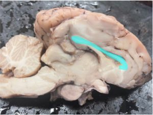
The Corpus Callosum
Revisit Cortical Brain Lobes, the Brainstem, and the Cerebellum
Hold and rotate your 1/2 brain specimen. Look at the medial view. Identify the lobes. Can you see them all? As aforementioned, you are not expected to know the precise boundaries of most lobes of the sheep brain; however, you should be able to identify their general vicinity and location relative to each other.
Identify the brainstem and the cerebellum.
Note the similarities and differences of the medial view of structures versus other views.
Test Yourself (or Each Other)
Identify the following structures on your specimen:
- corpus callosum
- cortex
- brainstem
- cerebellum
- general location of the frontal lobe
- general location of the parietal lobe
- general location of the temporal lobe
- general location of the occipital lobe
Name one function associated with each cortical lobe (Think back to material covered in class and Chapter 2).
How many frontal lobes are in each brain?
Is the corpus callosum best viewed from a more medial slice or a more lateral slice? Why?
Can you identify a brain structure covered in class and Chapter 2 but not specifically during this lab? Which one and where is it?
CLEAN UP
When you have finished, please rinse the dissection tray and clean your tools in the designated sink. If you are in an 8:30 am or a 10:00 am lab, please stack the trays in opposite directions (rather than sitting one inside of another) at the centre of your tables, and place your cleaned scissors, scalpels, probes, and/or forceps on a clean, dry paper towel. If you are in the 11:30 am lab, please stack the trays in opposite directions (rather than sitting one inside of another)at the back of the lab, and place your cleaned scissors, scalpels, probes, and/or forceps in the appropriate wooden tool box.
Lab Assignment Instructions
Lab Assignment Instructions
You should complete one of the two homework options given on Moodle.
First Option:
Moodle quiz
Second Option:
Knowing about the brain is important for personal health as well as to help make people good citizens!
Select one concept or fact from this lab session to teach as part of an Instagram post promoting a piece of knowledge you think people ought to know about the brain (think of this like a public service announcement) and/or the Antigonish Brain Bee. Incorporate a figure, photo, or video to teach about your fact/concept. To get the most of out this assignment, select a fact/concept that you are having trouble remembering/learning. See an example of an assignment on our Instagram account.
The post should be accurate; relevant to the material covered during this lab; at an appropriate level for a high school student, teacher, or parent audience; and have potential to generate interest in brain education and/or the Brain Bee. Be sure to submit an editable file or link to an editable file – which allows copying or editing. We recommend using tools such as PowerPoint or Canva. Also, be sure that you are permitted to share any content you use (e.g., clipart, music) by checking the copyright and/or license. This is an important point that often gets overlooked: Be sure not to include any images or content that is copyrighted, and be sure to include licensing information for all content you do use. (You could use your own photos or images.)
In addition to your post, include a document that reports your selected audience (high school students, teachers, or parents if you are advertising for the Brain Bee OR general public because you are giving a public service announcement), whether you permit the use of your post for advertising purposes for the upcoming Brain Bee (only useful for those post created for this purpose), and if so, which license you will apply to your work (.
Submit your assignment (the post and the document about permissions and license) on Moodle. See due date on Moodle.
Grading rubric (partial points will be awarded as appropriate)
| Criterion |
Below expectations
|
OK
|
Excellent
|
| Relevancy |
Not relevant to material covered in this lab
|
Partially relevant
|
Completely relevant
|
|
0 points
|
1 point
|
2 points
|
|
|
0 points
|
1 point
|
2 points
|
|
|
0 points
|
1 point
|
2 points
|
|
|
0 points
|
1 point
|
2 points
|
|
|
0 points
|
1 point
|
2 points
|
