22 Lab 4: Cellular Anatomy Lab (Neurons)
Erin Mazerolle and Sherry Neville-MacLean
Learning Objectives
Type your learning objectives here.
- Review cellular anatomy by investigating the components of a multipolar neuron smear through the use of a compound (or light) microscope
- Determine which parts of the neuron are (and are not) made visible using light microscopy.
- Construct a model of a multipolar neuron to strengthen knowledge of parts.
- Redesign model to account for types of neurons other than the multipolar neuron.
Tasks
One of the two lab tasks for this week involves using a microscope to examine a neuron smear. A neuron smear is a slide in which a neuron has be “smeared”.
Procedure for Neuron Smear Investigation
- Begin by using the alcohol and KimWipes at your station to clean the ocular lenses of the microscope. Place a bit of alcohol on a wipe, and work in increasingly smaller circles for each lens.
- Watch this short video to be sure you know how to focus the microscope. Although the presenter talks about the use of the 100X objective lens, it requires oil, and we will not be using this magnification. There is also a quick mention of adjusting the light level; given that we are working with a different brand of microscope, adjusting the level of light will be accomplished in a slightly different way. Your lab instructor can demonstrate if you need to adjust.
- Have a look through the lens to view the neuron using the 4X, 10X, and 40X levels of magnification objective lenses to determine which parts you can and cannot identify. (Note: the eyepiece also magnifies samples at 10X, so the resulting magnifications you will view at 40X, 100X, and 400X.)
- Have one group member record a short video with their phone while bringing the neuron into focus through one of the lenses. Use this video to capture an image in which the neuron appears clear. Post that photo and the markup you compose as your answer to question #2.
Note: Getting a photograph of a neuron smear: While some students may be able to quickly capture a photo of the neuron smear slide at a given magnification in such a way that they can use the photo to demonstrate different parts of the neuron, others will want to take a short video, and then, use the video to capture a still-frame photograph. - Answer the other Moodle questions related to your investigation of the neuron smear. You can use the labelled image below for comparison. Its only weakness is that the nodes of Ranvier are not labelled.
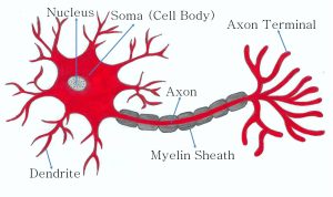
Source: https://commons.wikimedia.org/wiki/File:Neuron_typical_structure.jpg – License: CC-BY-SA-4.0
- Clean your microscope, unplug it, and cover it.
Building a model
Neuron modelling kits will be available and used for the second task. There are no instructional sheets. Instead, you use your knowledge of Chapter 3: Cellular Anatomy of Neurons to evaluate the modelling kit. Use the model(s) to complete the worksheet. While completing this task, 1) determine the strengths and weaknesses of the multipolar neuron model, and 2) reimagine the model to improve the weakness you identify. Do the same for the other types of neurons.
